

US-Based Nigerian Scholar Develops Low-Cost AI Tool for Brain Tumor Detection
US-Based Nigerian Scholar Develops Low-Cost AI Tool for Brain Tumor Detection
By Kunle Awosiyan
A United States-based Nigerian researcher, Oladele Ayomide Benjamen, has developed a low-cost artificial intelligence (AI) tool that could make brain tumor analysis more accessible, particularly in hospitals and research centres with limited computing resources.
Benjamen, a PhD student in Chemistry at Southern Methodist University, Texas, told our reporter that his research is focused on ensuring that advanced medical AI is not limited to wealthy institutions with expensive computing infrastructure.
“My research is helping make medical AI more accessible, so that hospitals and researchers with limited resources are not left behind,” he said.
His work comes at a time when AI is rapidly transforming cancer diagnosis. A 2025 systematic review and meta-analysis of 79 studies found that AI achieved an overall diagnostic accuracy of about 95.2 per cent in detecting and classifying brain tumors from MRI scans.
Unlike many existing AI systems that require costly high-performance computers equipped with Graphics Processing Units (GPUs), Benjamen’s research demonstrates that powerful brain tumor analysis can be performed using the Central Processing Unit (CPU) found in ordinary desktop and laptop computers.
Often described as the “brain” of a computer, the CPU executes software instructions and manages data processing. By designing AI models that run efficiently on CPUs, hospitals without expensive GPU hardware can still deploy advanced medical imaging tools.
His study, “Lightweight 3D U-Net for Brain Tumor Segmentation on CPUs: Enabling Deep Learning in Low-Resource Environments,” developed an efficient AI model capable of accurately identifying and mapping brain tumors while operating on standard computers.
“This is uncommon because most state-of-the-art AI models require powerful GPU-equipped systems for training and deployment,” Benjamen explained.
A graduate of Obafemi Awolowo University, Ile-Ife, where he earned a Bachelor’s degree in Chemistry, Benjamen previously worked as a Biomedical AI Engineer at the Medical Artificial Intelligence (MAI) Laboratory in Lagos and also held AI and bioinformatics roles at DycoVue and Genomac.
The CPU-based brain tumor project was carried out at MAI Lab alongside researchers including Charity Umoren, under the supervision of Dr. Maruf Adewole, Executive Director of MAI Lab, Prof. Fatade, Managing Director, and Prof. Udunna, Scientific Director at McGill University, Canada.
According to Benjamen, brain tumor diagnosis and treatment planning rely heavily on MRI scans, yet access to MRI facilities remains highly unequal. Across sub-Saharan Africa, there are fewer than one MRI scanner per one million people, making timely diagnosis difficult.
Even where MRI scans are available, many AI applications require expensive computing infrastructure, creating another barrier for hospitals in developing countries.
His research seeks to remove that obstacle by developing lightweight deep-learning systems that function effectively on conventional computers.
Beyond this work, Benjamen also contributed to MAPS-Glioma, an international research project involving Nigerian and Tanzanian scientists aimed at improving AI-assisted brain tumor analysis using medical imaging from resource-limited environments.
The project was part of the international BraTS-Africa Challenge, where the team ranked among the top-performing groups, providing independent recognition of the quality and relevance of their work.
Benjamen said his long-term ambition is to combine artificial intelligence, chemistry, medical imaging and nanotechnology to develop practical healthcare solutions.
“My long-term goal is to combine technology and science to create solutions that are not only innovative but useful. By bringing together AI, chemistry and healthcare research, I hope to develop tools that improve lives, support medical progress and solve real societal problems,” he said.
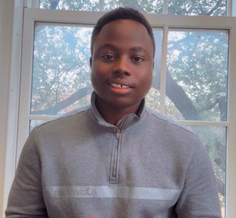
Now pursuing doctoral research in the United States, his work extends into nanochemistry, where he is investigating nanocluster-based platforms that could support future advances in cancer diagnosis, therapy and biomedical materials.
Commenting on the significance of the research, Raymond Confidence, Programme Chair of Spark Academy at McGill University, Canada, said the innovation addresses one of the biggest challenges facing healthcare systems in developing countries.
“For Nigeria and other resource-limited settings, the major benefit of this work is accessibility. It demonstrates that deep-learning tools for medical imaging can be trained and used on standard laptops and desktops, reducing dependence on costly infrastructure while supporting local capacity building in AI-driven healthcare,” he said.
Other studies have also demonstrated AI’s growing role in cancer diagnosis. A review published by the National Library of Medicine, titled Artificial Intelligence-Based Approaches for Brain Tumor Segmentation in MRI, concluded that AI enables faster and more accurate identification of brain tumors than traditional manual methods, improving diagnosis, treatment planning and clinical research.
Similarly, researchers at Mayo Clinic recently reported that an AI system can analyse routine pathology slides to help doctors classify meningiomas, the most common primary brain tumors in adults and estimate the likelihood of tumor recurrence.
Published in The Lancet Digital Health, the study showed that AI can extract valuable molecular information from standard pathology images, reducing reliance on expensive DNA methylation testing that remains unavailable in many hospitals.
Despite these advances, medical experts emphasise that AI is designed to support, not replace clinicians. Final diagnosis and treatment decisions continue to rest with qualified radiologists and pathologists.
Another major international breakthrough is Hetairos, an AI system developed in Germany that can classify brain tumors in approximately 12 minutes using tissue images.
Trained on more than 11,000 samples from 9,606 patients, the technology has demonstrated significant potential to accelerate diagnosis while remaining under clinical supervision.
As AI continues to reshape healthcare globally, Benjamen’s work highlights how innovation from Nigerian researchers can help bridge the technology gap and expand access to quality healthcare in underserved communities.